Description
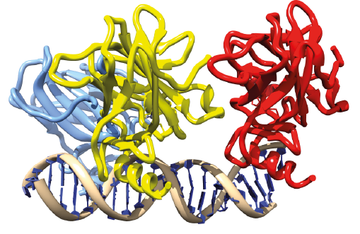
This class contains only 3 similar TFs, but it is one of the most well known because one of the TFs, TP53, is connected to a wide variety of cancers. Typically, this TF gets either silenced or its coding sequence is lost when cells undergo malignant transformation. All p53 TFs have a dedicated protein-protein interaction interface contained in a specific domain that causes them to form quadrimers even when occurring in free solution phase, besides this they have also a dimerization interface located in the DBD. Consistently with the layout of their multimerization interfaces, they bind DNA as 2+2 configurations recognizing duplexes of sites with RRCATGYY sequences with highest affinity for combination where the duplexes are separated by a 2bp spacer.
|
Structure
 |
